High-magnification imaging reveals the critical functional architecture of the central pore within drone bee cell caps. By clearly displaying the precise structure of these hard, conical silk caps, researchers obtain the microscopic physical evidence necessary to link these pores to essential biological functions, specifically gas exchange and chemical signal transmission.
Core Insight: The structural clarity provided by high-magnification systems validates the central pore as a deliberate biological adaptation, not a random artifact. It provides the missing anatomical link explaining how worker bees can monitor the health of sealed larvae and detect internal threats like parasites.
Unveiling the Micro-Architecture
Visualizing the Hard Cap Structure
High-magnification imaging systems allow for the detailed observation of the drone cell cap's composition.
These systems reveal that the caps are not simple covers but are hard, conical structures woven from silk.
The Formation of the Central Pore
The imaging confirms that the central pore is a specific feature formed by the drone larvae prior to pupation.
It is distinct from the surrounding silk matrix, serving as a dedicated channel between the internal cell environment and the outside world.
Linking Structure to Biological Function
Evidence for Gas Exchange
The microscopic verification of an open, unobstructed pore supports the theory of respiration.
The structure provides a physical pathway necessary for air circulation, allowing the developing pupa to exchange gases through the hard silk cap.
The Pathway for Chemical Signaling
Beyond respiration, the pore's architecture suggests a role in communication.
The imaging provides evidence that these openings act as conduits for chemical signals to exit the sealed cell.
Mechanisms of Disease Detection
This structural insight helps explain a critical hygiene behavior in the hive.
Researchers can now correlate the physical presence of the pore with the ability of worker bees to detect parasites or pathogens hiding within the cell.
The pore functions as a "scent window," allowing the chemical signatures of disease to reach the worker bees patrolling the comb.
Understanding the Limits of Structural Insight
Anatomy vs. Activity
While high-magnification imaging clarifies the structure of the pore, it does not visualize the active flow of gases or chemicals.
The images provide the anatomical proof that a pathway exists, but they must be paired with other data to measure the volume or type of transmission occurring.
Static Observation
The insights gained are primarily morphological.
The imaging captures the static state of the silk cap and pore, providing a snapshot of the physical machinery rather than the dynamic biological process in real-time.
Applying These Insights to Research
## Implications for Biological Study
- If your primary focus is functional anatomy: Use these imaging insights to define the precise geometry of the conical silk cap and how the larva constructs the pore before pupation.
- If your primary focus is colony pathology: Leverage the structural evidence of the pore to model how chemical signals regarding parasites travel from the larva to the worker bees.
High-magnification imaging transforms the central pore from a theoretical opening into a confirmed, functional interface for hive communication and health.
Summary Table:
| Feature Observed | Microscopic Insight | Biological Function |
|---|---|---|
| Cap Composition | Hard, conical silk structure | Structural protection & durability |
| Central Pore | Deliberate, unobstructed channel | Dedicated gas/chemical conduit |
| Silk Matrix | Woven larvae-formed silk | Physical barrier against external threats |
| Pore Architecture | "Scent window" design | Pathway for chemical signals & worker detection |
Scale Your Apiary Operations with HONESTBEE Precision Tools
At HONESTBEE, we understand that deep biological insights like the architecture of drone cell caps drive better beekeeping practices. Whether you are a commercial apiary looking to optimize colony health or a distributor seeking high-quality equipment, we provide the comprehensive wholesale solutions you need.
From advanced hive-making machinery and honey-filling systems to a full spectrum of beekeeping tools and essential consumables, our portfolio is designed to enhance efficiency and productivity. Let us help you integrate professional-grade hardware into your business to better manage hive pathology and honey production.
Ready to upgrade your equipment? Contact us today to explore our wholesale catalog!
References
- Gard W. Otis, Deborah R. Smith. Drone cell cappings of Asian cavity-nesting honey bees (Apis spp.). DOI: 10.1007/s13592-021-00864-8
This article is also based on technical information from HonestBee Knowledge Base .
Related Products
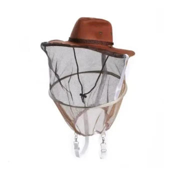
- Premium Cowboy Beekeeper Hat with Visibility Veil Outdoor Professional Beekeeping Protective Gear
- Square Folding Beekeeping Hat with Integrated Veil
- High-Definition Beekeeper Hat with Round Veil Mesh for Beekeeping
People Also Ask
- What protective gear is recommended for beekeeping? Essential Safety for Beekeepers
- What types of protective clothing and gear are available to beekeepers? Essential Safety Guide for Modern Apiarists
- How does beekeeping protective gear facilitate the queen bee selection process? Enhancing Precision in Queen Breeding
- What factors influence the choice of protective equipment used during honey harvesting? Essential Selection Guide
- What is the general protective gear recommendation for new beekeepers? Your Essential Beginner's Safety Guide