Compound microscopes are the definitive instrument for detailed honeybee analysis because they provide the high magnification and resolution necessary to visualize sliced or specially mounted tissue samples. Unlike stereo microscopes, which are limited to surface views, compound microscopes enable observation at the cellular level, allowing researchers to examine intricate microstructures such as sensory hairs on mouthparts or the barbs of a sting.
The shift from general observation to scientific rigor requires a compound microscope. It offers the micron-level precision needed to identify pathogens, measure specific anatomical features for taxonomy, and study physiological responses that are invisible to lower-power optics.
Unmatched Optical Capabilities
Superior Magnification and Resolution
To understand the physiology of a honeybee, you must go beyond the gross anatomy visible to the naked eye or a stereo microscope. Compound microscopes provide significantly higher magnification, which is essential for resolving fine details that would otherwise appear blurred or invisible.
Precise Micron-Level Measurement
Scientific taxonomy often relies on strict measurements of minute features. High-power biological microscopes provide the micron-level precision required to measure the length of hair roots and tips on the fifth abdominal tergite. They also allow for the clear definition of boundaries on tomentum bands (hair bands) found on the fourth tergite.
Advanced Lighting Techniques
Standard surface inspection is insufficient for internal analysis. Compound microscopes utilize high-power transmitted light, which shines through the specimen. This allows for the examination of internal structures within sliced tissues, rather than just the exterior surface.
Critical Applications in Research and Health
Pathological Diagnosis and Disease Control
The health of an apiary often depends on the early detection of microscopic threats. Laboratory-grade compound microscopes are standard for diagnosing specific pathogens, including viruses, bacteria, fungi, and mites.
Targeted Epidemic Prevention
By accurately identifying the specific agent causing a disease, technicians can formulate targeted control plans. This precise pathological analysis is the core technical method used to inhibit the spread of epidemics within a bee colony.
Physiological and Toxicological Studies
For advanced research, such as studying how bees respond to toxins, researchers must look at the cellular level. Compound microscopes allow for the observation of toxicological responses within the tissue, providing insights that macroscopic observation cannot yield.
Understanding the Operational Requirements
Sample Preparation is Mandatory
The primary trade-off when using a compound microscope is the requirement for laborious sample preparation. Because the light must pass through the object, you cannot simply place a whole bee under the lens.
The Necessity of Mounting
Specimens must be sliced thin or specially mounted on glass slides to be viable. This makes the compound microscope a tool for the laboratory, not for quick, non-destructive field inspections of live insects.
Making the Right Choice for Your Goal
To maximize the value of your research, match your objective to the specific capabilities of the instrument:
- If your primary focus is Academic Taxonomy: You need a compound microscope to measure hair length and tomentum width with micron-level accuracy for species identification.
- If your primary focus is Disease Management: You must use this tool to visually confirm the presence of specific microscopic pathogens like bacteria or fungi to select the correct treatment.
- If your primary focus is Advanced Physiology: Rely on high-magnification transmitted light to observe cellular structures and sensory organs on prepared tissue slides.
Precision in analysis leads to precision in understanding, ensuring the health and proper classification of the colony.
Summary Table:
| Feature | Compound Microscope Capability | Benefit for Bee Research |
|---|---|---|
| Magnification | High (Up to 1000x+) | Visualizes cellular structures & pathogens |
| Light Source | Transmitted Light | Examines internal tissues & sliced samples |
| Precision | Micron-level | Accurate measurement of hairs & tomentum bands |
| Application | Pathological Diagnosis | Identifies bacteria, fungi, and viruses |
| Sample Type | Sliced/Mounted Slides | Detailed study of mouthparts, stings, and organs |
Elevate Your Apiary's Scientific Standards with HONESTBEE
At HONESTBEE, we understand that commercial success in beekeeping requires more than just field experience; it demands scientific precision. We cater to commercial apiaries and global distributors by providing a comprehensive wholesale offering of professional-grade beekeeping tools and laboratory-ready machinery.
Whether you are setting up a diagnostic lab to prevent epidemics or scaling your production with our honey-filling machines, our portfolio covers the full spectrum of industry essentials—from specialized hardware to honey-themed cultural merchandise.
Ready to scale your operations with premium equipment? Contact us today to explore our wholesale solutions and see how our expertise can drive your business forward.
References
- Daniel Basterfield. The Future Of Beekeeping?. DOI: 10.1080/0005772x.2011.11417399
This article is also based on technical information from HonestBee Knowledge Base .
Related Products
- HONESTBEE Professional Mini J-Hook Hive Tool for Beekeeping
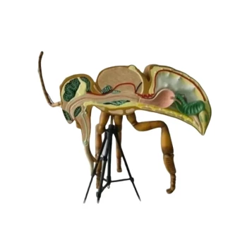
- HONESTBEE Anatomy Bee Model Detailed Anatomical Display for Education and Study
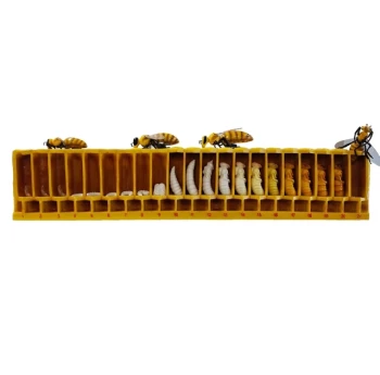
- Honey Bee Lifecycle Model: A Detailed Honeycomb Display of Bee Development
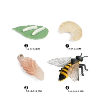
- Honey Bee Life Cycle Model 4 Stage Educational Set for Kids Learning
- Economy Galvanized Beekeeping Honey Bee Smoker for Wholesale
People Also Ask
- How is a J-hook hive tool designed and what is its primary use? Master Effortless Hive Inspections
- What is a J-hook hive tool and why is it favored by some beekeepers? Master Gentle Frame Handling
- What is the tool used in beekeeping? Master the 4 Essential Tools for Your Hive
- What is the importance of using the right tools in honey production? Boost Quality, Yield & Profitability
- What specific challenges do beekeeping hive tools address during hive maintenance? Essential Solutions for Apiaries