High-magnification laboratory microscopes serve as the definitive verification tool for diagnosing internal honey bee ailments that are impossible to detect through visual inspection alone. Their primary function is to enable the precise identification and quantification of microscopic parasites, specifically Nosema spores and tracheal mites (Acarapis woodi), by examining dissected bee abdomens and respiratory systems.
The Core Insight While external symptoms can suggest a colony is unwell, high-magnification microscopy provides the pathological certainty required for effective treatment. It transforms a general diagnosis of "failure to thrive" into a confirmed identification of specific pathogens, forming the technical foundation for targeted disease control.
The Mechanics of Microscopic Diagnosis
Beyond External Observation
A standard inspection or use of a 50X magnifying glass allows for the assessment of external symptoms, such as deformed wings or abdominal swelling. However, these tools cannot reveal the root cause of internal infections.
Deep Tissue Analysis
High-magnification microscopes (typically 400X) allow technicians to look inside the specimen. By analyzing abdominal homogenates or tracheal sections, the microscope reveals pathogens residing within the honey bee's internal organs.
Quantitative Assessment
This technology does not just confirm presence; it allows for qualitative analysis. Technicians can count spore loads or assess the severity of a mite infestation, providing data on the intensity of the disease.
Targeting Specific Pathogens
Identifying Nosema Spores
One of the primary applications is the detection of Nosemosis. Technicians use the microscope to identify Nosema spores based on their distinct morphology within abdominal fluid samples.
Detecting Tracheal Mites
The microscope is essential for locating Acarapis woodi. By examining the tracheal tubes (the bee's respiratory system), the microscope reveals these microscopic mites that would otherwise go unnoticed until the colony collapses.
Enabling Strategic Action
This precise identification is the "core technical method" for apiary management. Once the specific pathogen—whether fungus, mite, or bacteria—is identified, beekeepers can formulate targeted prevention plans to inhibit the spread of the epidemic.
Understanding the Trade-offs
Sample Destruction
Unlike a 50X hand lens which allows for non-destructive inspection of live bees, high-magnification analysis requires dissection. This process involves sacrificing a sample of bees to protect the larger colony.
Limitation Regarding Viruses
While microscopes are excellent for parasites and fungi, they have limitations with smaller pathogens. Physical observation often serves as a complement to molecular detection methods when diagnosing complex viral or bacterial infections.
Making the Right Choice for Your Goal
To manage colony health effectively, you must match the diagnostic tool to the specific depth of analysis required.
- If your primary focus is rapid field assessment: Rely on a 50X high-magnification glass to spot external deformities, abdominal swelling, or body color changes without harming the bees.
- If your primary focus is confirming internal parasites: Utilize a 400X biological microscope to analyze abdominal and tracheal dissections for a definitive count of Nosema spores or tracheal mites.
True apiary health relies on moving beyond assumptions to evidence-based intervention.
Summary Table:
| Diagnostic Tool | Magnification | Primary Use Case | Sample Impact | Pathogens Detected |
|---|---|---|---|---|
| Hand Lens/Magnifier | Up to 50X | Rapid field assessment of external symptoms | Non-destructive (Live bees) | Deformed wings, body color, swelling |
| Biological Microscope | 400X and above | Definitive identification of internal parasites | Destructive (Requires dissection) | Nosema spores, Tracheal mites (Acarapis woodi) |
Secure Your Apiary’s Health with Professional-Grade Solutions
At HONESTBEE, we understand that accurate diagnosis is the first step toward a thriving colony. Whether you are a commercial apiary needing advanced machinery or a distributor looking to supply high-precision beekeeping tools, we provide the comprehensive wholesale support you need.
Why Partner with HONESTBEE?
- Precision Equipment: From high-performance honey-filling machines to essential hive-making hardware.
- Industry Expertise: We supply the full spectrum of beekeeping tools and specialized consumables tailored for large-scale operations.
- Targeted Value: We help you move beyond assumptions with equipment designed for pathological certainty and operational efficiency.
Contact HONESTBEE Today to explore our full product portfolio and elevate your beekeeping standards.
References
- Fekadie, Bereket. Studying Causes of Honey Bee Mass Death in Western Amhara Region, Ethiopia. DOI: 10.5281/zenodo.17948671
This article is also based on technical information from HonestBee Knowledge Base .
Related Products
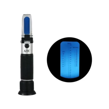
- Precision Honey Refractometer Instrument for Quality Assessment
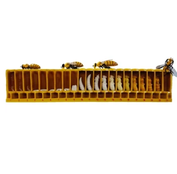
- Honey Bee Lifecycle Model: A Detailed Honeycomb Display of Bee Development
People Also Ask
- What are two critical factors for obtaining accurate results with a honey refractometer? Master Honey Testing Today
- How is the nutrient grade of honey measured? Master Brix and Moisture for Premium Quality Honey
- What is the purpose of a honey refractometer? Ensure Perfect Moisture for Harvest & Sales
- How is honey applied to the refractometer? Ensure Accurate Moisture Content Measurement
- How do professional scientific instruments contribute to honey quality testing? Ensure Purity and Market Compliance