High-power biological microscopy is the definitive standard for physically identifying and quantifying parasitic infections in honey bee colonies. Utilizing 400x magnification, this equipment allows technicians to perform direct visual screenings of bee tissues to diagnose specific threats like Nosema and tracheal mites.
While molecular methods are ideal for viruses, the optical microscope remains the essential tool for observing the physical morphology of larger parasites. It provides the only means to visually verify and count parasite loads within specific anatomical sections of the bee.
The Mechanics of Microscopic Screening
Diagnosing Nosema via Abdominal Homogenates
To screen for Nosema, technicians utilize the microscope to examine abdominal homogenates. This involves processing the abdomen of the bee into a liquid suspension.
Under 400x magnification, the observer scans this fluid for specific biological markers. The diagnosis relies on identifying Nosema based on its distinct spore morphology.
This process is not just binary (positive/negative); it allows for a quantitative assessment. By counting the visible spores, technicians can estimate the severity of the infection within the colony.
Identifying Tracheal Mites in Respiratory Sections
The screening process for tracheal mites (Acarapis woodi) requires a different preparation technique focused on the respiratory system.
Technicians prepare tracheal sections, slicing through the bee's thorax to expose the main breathing tubes. The high-power microscope is then used to peer directly inside these tubes.
The goal is to visually confirm the presence of mites residing within the trachea. This direct observation is the primary method for diagnosing this specific respiratory infestation.
Complementing Molecular Diagnostics
Microscopy does not replace advanced laboratory techniques; it partners with them.
While molecular detection methods are necessary for identifying invisible threats like bacteria and viruses, they are often overkill or less practical for macro-parasites. The high-power microscope serves as the physical validation layer in a complete diagnostic toolkit.
Understanding the Trade-offs
Manual Precision vs. Automation
Microscopic screening is labor-intensive compared to automated molecular tests. It requires the manual preparation of slides—either homogenizing abdomens or slicing tracheal sections.
Reliance on Technician Skill
The accuracy of this method depends heavily on the operator's ability to recognize morphology.
Distinguishing a Nosema spore from other debris, or identifying a mite within a complex tracheal section, requires a trained eye. Misidentification can lead to false negatives or incorrect spore counts.
Making the Right Choice for Your Diagnostic Goals
Microscopy is a versatile tool, but your specific protocol depends on the target parasite.
- If your primary focus is [Quantifying Nosema Infection]: Focus on preparing abdominal homogenates to perform accurate spore counts based on visual morphology.
- If your primary focus is [Detecting Tracheal Mites]: Prioritize the careful sectioning of the thoracic region to expose the tracheal tubes for direct internal inspection.
By mastering these visual screening techniques, you ensure a robust defense against the parasites that threaten colony health.
Summary Table:
| Parasite Target | Tissue Preparation | Magnification | Key Diagnostic Feature |
|---|---|---|---|
| Nosema Spores | Abdominal Homogenate | 400x | Distinctive spore morphology & quantitative counts |
| Tracheal Mites | Thoracic Tracheal Sections | 400x | Direct visual identification of mites inside breathing tubes |
| Viruses/Bacteria | Molecular/PCR (Non-Microscope) | N/A | Genetic markers (Microscopy serves as a physical validation) |
Scale Your Colony Health Diagnostics with HONESTBEE
Maintaining a healthy apiary starts with the right diagnostic tools. At HONESTBEE, we empower commercial apiaries and global distributors by supplying a comprehensive range of professional beekeeping machinery, specialized hardware, and essential industry consumables.
Whether you are setting up a laboratory for parasite screening or optimizing your honey production with our advanced filling machines, we provide the industrial-grade equipment needed to ensure colony vitality and business growth.
Ready to upgrade your wholesale beekeeping supply chain?
Contact HONESTBEE Today to discuss how our tools and machines can enhance your operations.
References
- Félicien Amakpe, Dirk C. de Graaf. Discovery of Lake Sinai virus and an unusual strain of acute bee paralysis virus in West African apiaries. DOI: 10.1007/s13592-015-0372-z
This article is also based on technical information from HonestBee Knowledge Base .
Related Products
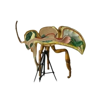
- HONESTBEE Anatomy Bee Model Detailed Anatomical Display for Education and Study
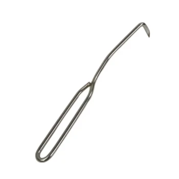
- Precision Stainless Steel Frame Cleaner for Hive Grooves and Corners
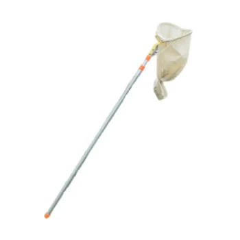
- HONESTBEE Professional Telescopic Pole Bee Swarm Catcher
- HONESTBEE Adjustable Voltage Wire Embedder with Digital Display
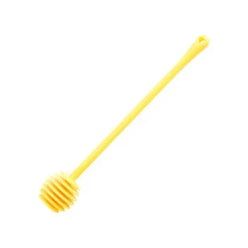
- Professional Plastic Honey Dipper for Easy Honey Drizzling
People Also Ask
- What criteria should be considered in the selection process for honey bee breeding? Top Traits for Sustainable Apiaries
- Are honey bees good to have around? The Essential Role and Ecological Impact of Bees
- Why must strict sanitary and health prevention protocols be implemented for honeybee transport? Maximize Colony Survival
- Why is maintaining specific relative humidity levels critical in caged honey bee experiments? Ensure Research Validity
- What should be considered when learning the behavior of honey bees and their queens? Master Colony Management